News Center
If you have tried to conceive for twelve months under age 35, six months at 35 or older, or any length of time past 40, the watch-and-wait phase has already ended by guideline standards. Three body signals deserve a US fertility clinic visit sooner: a luteal phase that keeps shrinking, one early miscarriage, and a "normal" semen report that conceals DNA damage. Each one changes what IVF can still accomplish.
Fertility medicine has a formal clock. The American Society for Reproductive Medicine (ASRM) defines infertility in its committee opinion published in Fertility and Sterility (2017 revision) as failure to conceive after twelve months of regular unprotected intercourse for women under 35, after six months for women aged 35 to 40, and it recommends immediate evaluation for women over 40. Those thresholds are the calendar part of the answer. The five signs below are the body part, and three of them routinely get misread by patients and general gynecologists alike:
Signs four and five are straightforward, and most people eventually recognize them on their own. Signs one through three are different. Each carries a specific mechanism, a specific misreading, and a specific set of actions that a reproductive endocrinologist handles very differently from a general obstetrics practice. The rest of this article stays on that line: what the signal is, why it gets misread, and what to actually do about it.
The menstrual cycle has two halves. The follicular phase runs from the first day of bleeding to ovulation and varies widely in length between women and between cycles. The luteal phase runs from ovulation to the next period. In a healthy ovulatory cycle it stays remarkably stable, typically twelve to fourteen days, because it is governed by the lifespan of the corpus luteum, the temporary gland that produces progesterone after ovulation. This stability is what makes the luteal phase a diagnostic window. When it begins to compress, the most common explanation is declining ovarian function: fewer responsive follicles, weaker preovulatory estradiol signals, a corpus luteum that forms inadequately and quits early.
Here is the part that creates false reassurance. Because the follicular phase absorbs most of the variability, a woman whose cycles shorten from 30 days to 26 days may see nothing alarming at all. Her period still arrives "on schedule." What has actually changed is that the follicular half shrank and the luteal half quietly lost days. Many reproductive specialists consider a luteal phase consistently under ten days worth investigating, because implantation depends on progesterone support lasting long enough for the embryo to embed and signal. A corpus luteum that fails at day eight leaves an embryo trying to implant into a lining that is already being shed.
Flow volume adds a second clue. Lighter bleeding over the same number of days often reflects a thinner endometrium, which tracks with the same underlying trend. A period that once needed heavy protection for three days and now passes with two light days, cycle after cycle, is data. It is data that a busy fifteen-minute annual exam almost never captures, because nobody asks the patient to count days since ovulation.
The misreading has an institutional history worth understanding. The American College of Obstetricians and Gynecologists (ACOG), in Committee Opinion No. 541 on the diagnosis of luteal phase defect (2012), concluded that the evidence does not support "luteal phase defect" as a standalone diagnosis and discouraged routine endometrial biopsy for it. That position is scientifically defensible for isolated cases, and it is one reason general practitioners often dismiss the complaint entirely. The gap sits between two facts: the diagnosis label is weak, yet the trend of a persistently short luteal phase combined with falling flow is a real signal of waning ovarian reserve that deserves testing rather than reassurance. Many clinicians describe luteal decline as appearing one to two years before cycles become visibly irregular, which makes it the earliest and least-acted-upon sign on this list.
A common counterargument deserves space: perimenopause also shortens luteal phases and lightens flow, so how do you tell the difference at 38 or 40? The honest answer is that symptoms overlap, and that ambiguity is itself a reason for the clinic visit rather than against it. AMH and AFC results, interpreted alongside age, separate "ovarian reserve running low, IVF timeline matters now" from "normal age-related transition." Guessing wrong in either direction costs time, and time is the one input IVF cannot manufacture.
Roughly 80 percent of miscarriages occur in the first trimester, according to ACOG patient guidance, and chromosome abnormalities in the embryo account for about half of those first-trimester losses. Professional societies, including ASRM and the European Society of Human Reproduction and Embryology (ESHRE), define recurrent pregnancy loss as two or more clinical pregnancy losses, and that definition drives standard practice: after one loss, most patients are told the event was likely random, to grieve, and to try again. For a 28-year-old who conceived within three months, that advice is statistically reasonable. The same sentence delivered to a 38-year-old who needed eleven months to conceive is where the misreading begins.
The statistics behind the difference are well documented. Age-specific miscarriage risk rises steeply: broadly cited figures from cohort studies place loss risk near 10 percent for women in their twenties, rising through the thirties and approaching 40 to 50 percent by the mid-forties. Meanwhile, time to conception lengthens with age for the same biological reason, because the proportion of chromosomally abnormal eggs climbs. Put together, a single early loss in a woman over 35 carries information on two fronts. It confirms that conception happened, which is genuinely good news about tubal patency and ovulation. It also documents, in the most concrete way possible, that an embryo with the chromosome profile of her remaining egg pool failed to develop. The next egg drawn from the same pool carries a comparable risk profile.
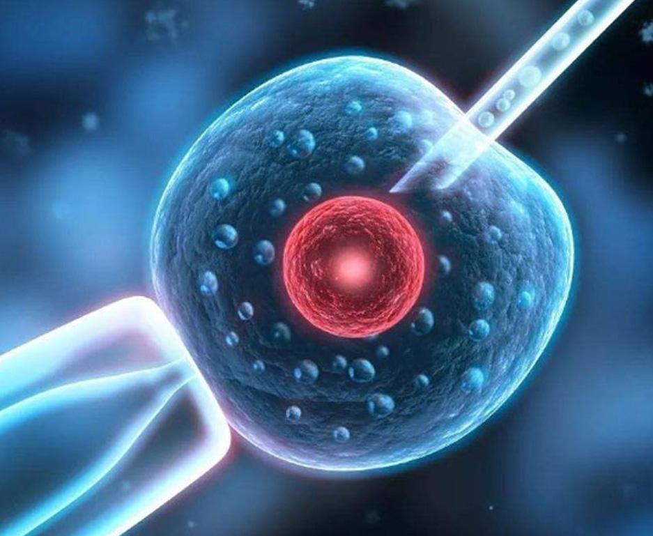
What general gynecology rarely volunteers at this point is that in vitro fertilization with preimplantation genetic testing for aneuploidy (PGT-A) exists precisely for this situation. PGT-A counts the chromosome sets in embryos created through IVF before transfer, allowing the selection of euploid embryos. For a patient whose losses are driven by egg aneuploidy, transferring a tested embryo addresses the dominant failure mechanism directly. It is worth stating plainly what PGT-A does and does not do: it reduces the chance of transferring an aneuploid embryo, it does not guarantee implantation, and it does not screen every possible genetic condition. Mosaic results complicate interpretation. Those caveats belong in a conversation with a reproductive endocrinologist, which is exactly the conversation that tends to happen eighteen months later than it should when everyone accepts "one loss is just bad luck" as the final word.
The boundary condition cuts the other way too, and it matters. PGT-A involves ovarian stimulation, egg retrieval, and embryo creation, which carries real cost, physical burden, and emotional weight. A single loss does not obligate anyone to IVF, and no credible physician frames it that way. The claim here is narrower: one loss plus advanced age plus slow conception is a legitimate medical indication to have the IVF conversation now, and the default of waiting another year quietly consumes the very resource, oocytes, that the treatment depends on.
Male factors contribute to a large share of infertility cases; commonly cited estimates attribute roughly 40 to 50 percent of cases, alone or combined, to the male partner. Despite that, the male workup often stops at one semen analysis, and when the report says "normal," the file closes. The phrase deserves unpacking.
The reference values come from the World Health Organization laboratory manual, most recently the sixth edition published in 2021. The 5th-percentile reference limits include a semen volume of 1.4 mL, sperm concentration of 16 million per mL, total sperm number of 39 million, progressive motility of 30 percent, and normal morphology of 4 percent. Two facts about these numbers change how they should be read. First, they describe the lower edge of the distribution among men in couples who conceived within twelve months, so a result above the limit indicates a probability position, never a guarantee. Second, a result below one limit does not equal sterility. "Within normal range" therefore means "not obviously at the bottom of the fertile distribution," which is a much weaker statement than patients usually assume.
The deeper gap is what the standard panel never measures at all. Sperm DNA fragmentation (DFI) reflects the integrity of the genetic material the sperm carries, and elevated fragmentation is associated with reduced fertilization, impaired embryo development, higher miscarriage rates, and unexplained infertility. DFI appears in no routine physical, no standard semen report, and often no fertility workup unless someone specifically orders it. The ASRM's Society for Male Reproduction and Urology published guidance (2023) noting that sperm DNA fragmentation testing may be considered in specific situations, including unexplained infertility and recurrent pregnancy loss. A couple can spend a year inside the system with a "normal" male workup while the relevant abnormality sits in a test nobody ran.
A realistic example shows the stakes. A couple in their mid-thirties tries for fourteen months. Her workup: regular ovulation, open tubes, adequate reserve. His single analysis: concentration of 22 million per mL, motility 45 percent, morphology 5 percent, all above WHO limits, stamped normal. Nobody orders DFI. Had it been ordered and returned elevated, the conversation would shift immediately toward IVF with ICSI and sperm selection rather than another year of timed intercourse. The report that ended the investigation was the very thing that should have extended it.
| WHO 6th ed. (2021) parameter | 5th-percentile reference limit | What it does not tell you |
|---|---|---|
| Semen volume | 1.4 mL | Nothing about DNA integrity |
| Sperm concentration | 16 million/mL | Whether sperm fertilizes an egg normally |
| Progressive motility | 30% | Whether the DNA inside is fragmented |
| Normal morphology | 4% | Function; shape correlates loosely at best |
Sign four, the guideline clock, needs only restating: twelve months under 35, six months at 35 to 40, immediate evaluation past 40, per the ASRM definition. Many patients wait longer than this because the calendar feels arbitrary when nothing "feels wrong." It is not arbitrary; it encodes the declining probability of spontaneous conception per cycle.
Sign five covers known diagnoses and failed treatments. Endometriosis, even surgically confirmed mild disease, erodes ovarian reserve over time and distorts the pelvic environment. Fibroids that indent the uterine cavity reduce implantation odds until removed. Anyone who has completed two or three IUI cycles without pregnancy has already generated outcome data: the treatment tier below IVF has been tested in her specific body and did not work. Continuing the same tier for another year repeats an experiment whose result is known. In all these cases the decision about IVF is less about whether and more about scheduling, and scheduling questions belong in a reproductive endocrinology office.
Patients acting on the three signals above are usually early in the process, so the clinic question is less about prestige and more about fit. Three organizational models cover most needs, and naming real examples helps anchor the search.
Large national networks suit patients who value geographic flexibility and standardized protocols. Shady Grove Fertility, headquartered in Maryland with more than 40 locations across the United States, operates on this model and has long served patients who may relocate or who want local monitoring with a larger center handling the procedure itself. HRC Fertility runs multiple sites across California and functions similarly for West Coast patients, with the density of Southern California locations reducing travel for monitoring appointments, which typically require early-morning visits every one to two days during stimulation.
High-volume, value-oriented centers fit patients paying out of pocket who prioritize cycle cost and transparency over amenity. CNY Fertility, based in New York State with several branch locations, is known for a high-volume, lower-price structure and publishes its pricing openly. The tradeoff involves less per-patient hand-holding and more self-driven logistics, which some patients manage well and others find stressful mid-cycle.
Whichever model fits, verify outcomes the same way for all of them. The CDC's ART reporting system and SART publish per-clinic success rates by age group and cycle type. Read those tables for your own age bracket, ask each clinic how it selects patients for transfer, and treat any single headline number, including favorable ones, as incomplete without that context. Clinic-published figures should always be checked against the official registry data rather than taken from marketing pages.
One final practical note. The first appointment at a US fertility clinic is a consultation plus a review of existing records, and it demands far less preparation than patients fear. Bring your cycle logs with luteal phase counts, the pathology or chromosome report from any loss, both copies of any semen analyses, and a written list of questions including the two that matter most here: whether IVF with PGT-A is indicated in this specific situation, and whether sperm DNA fragmentation testing should be added to the male workup. A single visit converts a year of ambiguous signals into a concrete plan, and unlike the eggs it protects, the appointment itself takes about an hour.